Read about the conditions that come along with injuries and sports injuries. See our approach to fixing your problems with our cutting edge solutions.

Concussions and Chiropractic

Concussions and Chiropractic

From car accidents to sports injuries, there are a number of possible causes of concussions. According to the CDC, a concussion is a type of traumatic brain injury that can affect the way the brain works. Although the severity of each one will vary, they all should be taken seriously. A few examples of some symptoms that may be associated with concussions include dizziness, headache, blurry vision, memory loss, nausea and vomiting. Thanks to improvements in technology, more concussions are being diagnosed than ever before and, as a result, fewer of them are being left untreated.

Not everyone may be aware of this fact, but chiropractors receive extensive training when it comes to recognizing concussions. Along with the actual head injury itself, there can often be a neck injury inflicted at the same time with symptoms of its own that may be difficult to differentiate from those of the concussion. Your chiropractor can not only help identify these injuries, but he or she will also help develop a treatment plan to alleviate painful symptoms and help your body heal properly! Sidney Crosby of the Pittsburgh Penguins is an excellent example of an athlete who received successful chiropractic treatment that helped improve both his cognitive and physical abilities after suffering a concussion during a hockey game.

If you ever suffer a head injury while playing a sport, be sure not ignore it and keep playing. Let a medical professional examine you to determine whether or not you have a concussion.

IT Band Syndrome treatment San Diego

Iliotibial Band Syndrome

Check out our updated post on IT band treatment found here: New Methods in Treating IT Band Syndrome!

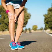
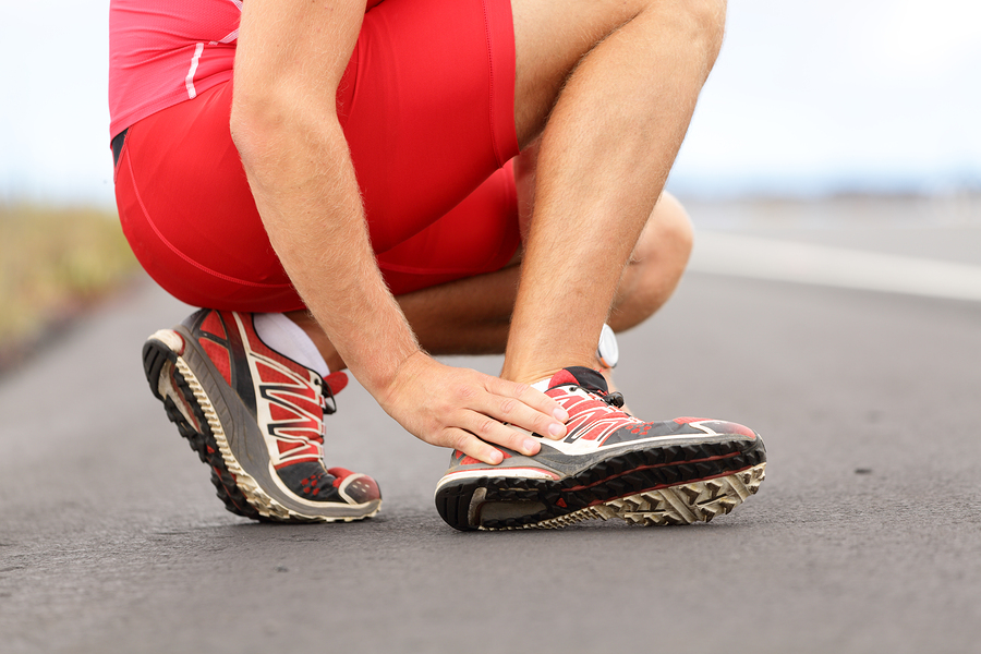
Iliotibial band syndrome (ITBS) is a painful, frustrating condition that affects the outside part of the knee. The IT band is a thickening of inelastic connective tissue which arises from the Tensor Fascia Latae and the Gluteal muscles and inserts into the outside of the tibia and Tibialis Anterior.1 During movement, the IT band approximates the lateral aspect of the knee, creating an “impingement zone” and irritates the tissues found there, primarily a highly innervated fat pad.2,5 With repetitive flexion and extension of the knee, particularly during excessive running, ITBS can develop and cause moderate to severe pain on the outside of the knee.

Who is at risk?

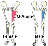
Active individuals are prone to iliotibial band syndrome due to chronic “overuse” type of trauma to the IT band and muscles of the thigh/buttock. A number of risk factors put a person at a greater risk for developing ITBS which include: training intensity/duration, running down hills, wearing shoes with poor arch support, biomechanical factors such as flat feet, gluteus medius weakness, etc.4  Runners who run while fatigued, may be at more of a risk for developing ITBS due to increased flexion at foot-strike; this results in greater impingement at the “impingement zone” of the knee leading to greater tissue irritation.2

Examination

Sports chiropractors will carefully evaluate the knee including: ROM, various orthopedic tests, and a specific palpatory assessment, which allows the practitioner to identify muscles that have developed myofascial adhesions, causing muscle tension and pain.

The knee is caught between two other main joints of the lower extremity: the ankle, and the hip. The IT band connects to the hip musculature and also has fascial connections to the tibialis anterior, the bulky muscle in the front of the lower leg.1 Examination of the hip, lower leg, and ankle may also be assessed in an attempt to determine dysfunctional muscles, joints, and structures which may be causing excessive stress at the outside of the knee.

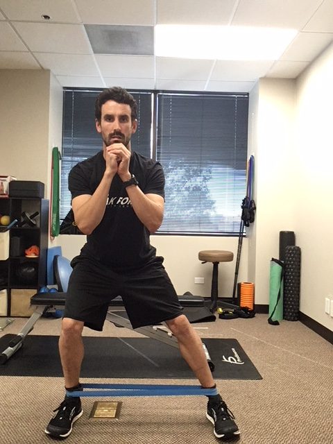
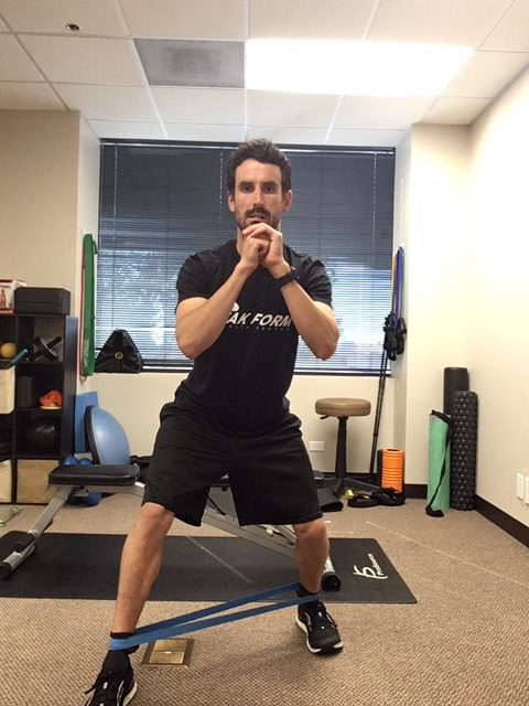
A functional evaluation will also be performed to determine movement patterns that may cause excessive stress on the knee/IT band. A gait assessment both walking and running may reveal over pronation at the foot, or hip dropping during the stance phase of the gait cycle. Squats and lunges may reveal deviation of the knee towards midline, which may indicate glute medius weakness on the same side.

Treatment

Iliotibial Band Syndrome Treatment Graston San Diego

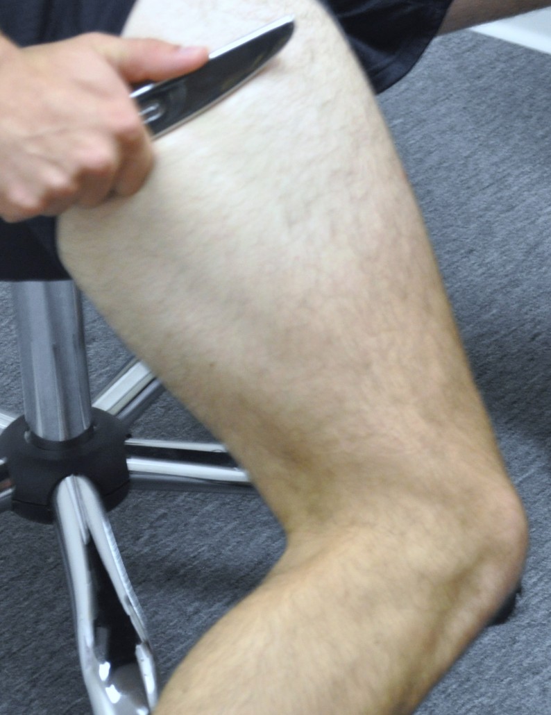
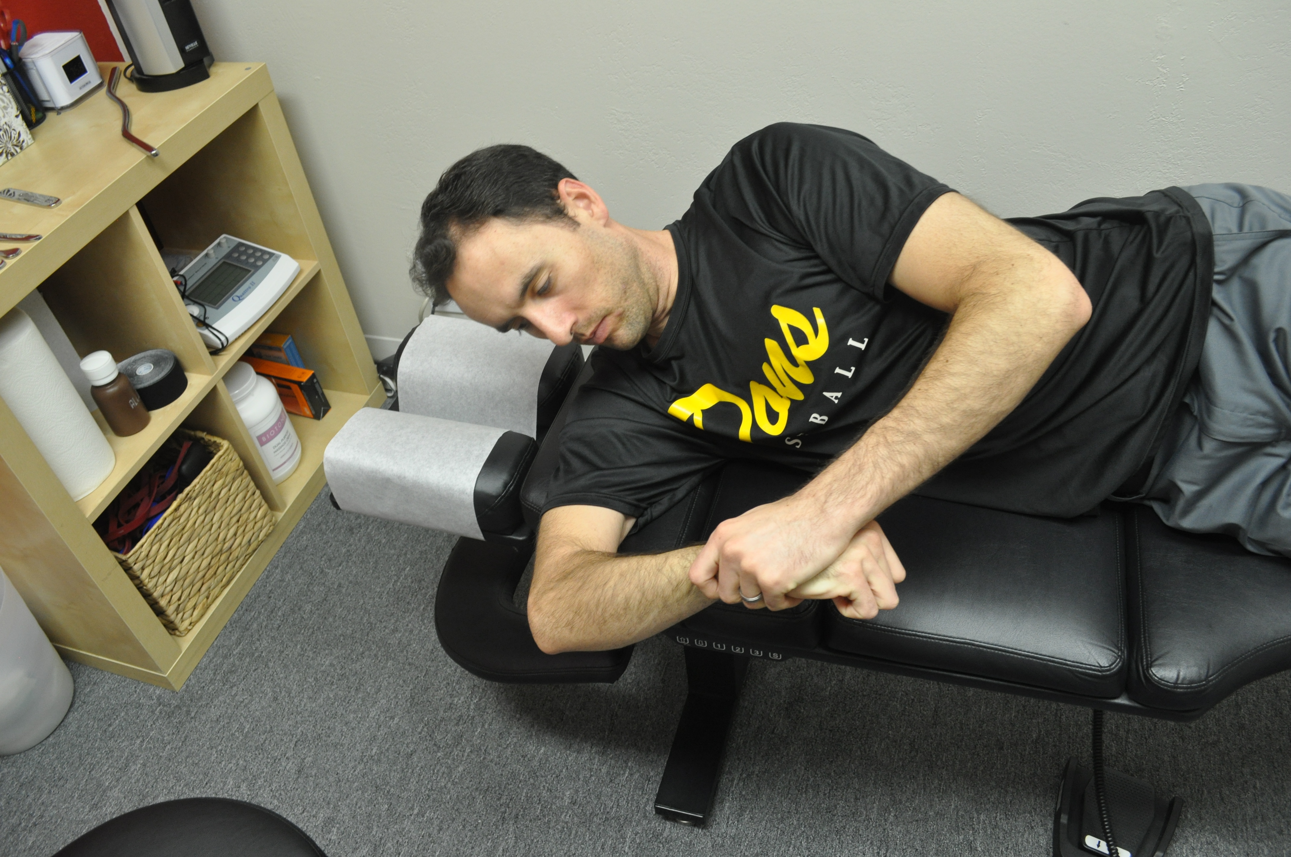
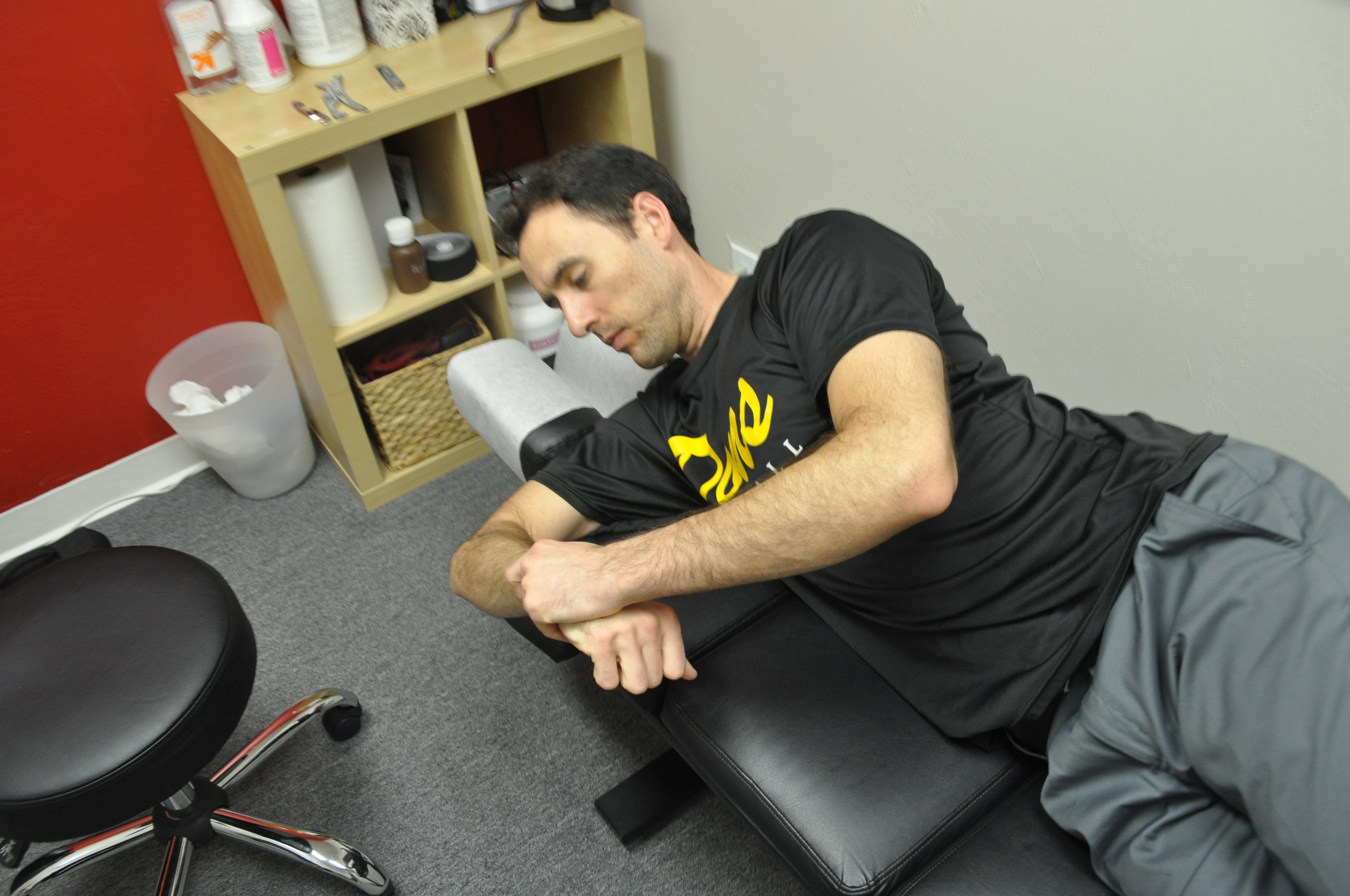
Graston® Technique applied to IT band/Quadricep.

Treatment depends on the stage at which the ITBS is found at the time of examination. To combat swelling and pain associated with ITBS, treatment primarily involves ice and modification of activity.3,4 Patients can ice the knee at home using ice massage for 3-8 minutes, two times per day to combat the inflammatory process.

Sports chiropractic care primarily involves addressing the soft tissue structures that affect the IT band. Active Release Technique (ART®) is used to remove myofascial adhesions (scar tissue) that have developed in the muscles that surround or connect into the IT band: gluteus maximus/medius, TFL, vastus lateralis (outside quadriceps muscle), biceps femoris (outside hamstring), and in some cases the tibialis anterior. The practitioner will have you perform specific movements for each structure involved, while deep pressure is applied to the structure to help stretch the myofascial adhesions to help restore proper function to the muscles and remove excessive tension that may be exacerbating the ITBS.

Some ART® protocols are designed to break down adhesions within the muscles, while other protocols are designed to restore relative motion between myofascial structures. The latter is important for IT band syndrome as the band itself lies over the large vastus lateralis and biceps femoris. Improving the movement between these structures will help remove tension at the outside of the knee.

Chiropractic adjustments may be used to improve the joint function of the low back, pelvis, hip, knee, and ankle to help improve the symptoms at the knee.

Rehabilitation

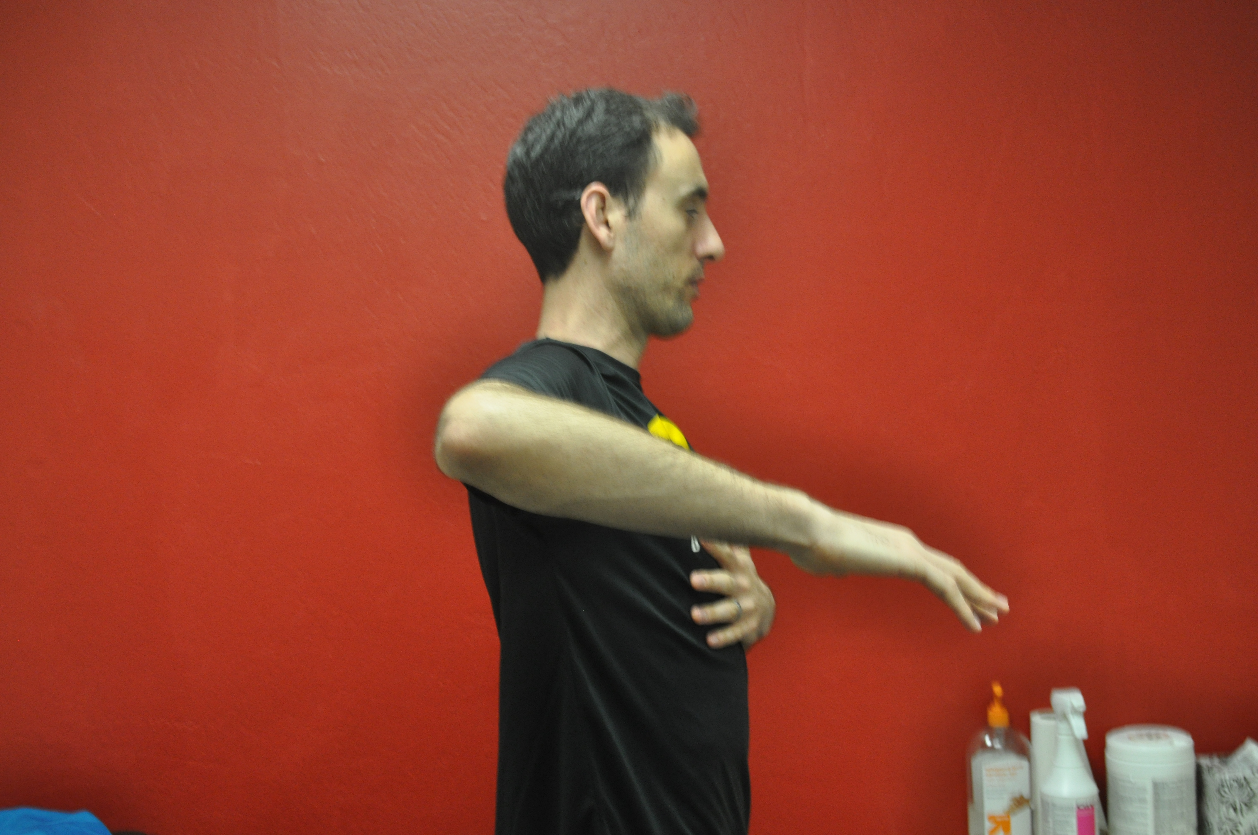
Once pain has significantly decreased, specific exercise protocols will be given to help prevent recurrence of symptoms and strengthen weak muscles such as the gluteus medius muscle. Gluteus medius weakness results in greater adduction angle at the knee (knee moves inward) during gait which results in over stabilization of the IT band leading to ITBS, trigger points, and contracted muscles.6,7 Proper strengthening of the glute medius will result in greater hip stabilization and keep the knee from deviating towards midline during dynamic movements (running, squatting, jumping).The IT band is an inflexible structure, however, IT band stretching can help relieve tension in the muscles that insert into the IT band. In addition to static stretching, the use of ischemic compression through the use of a foam roller can be used to successfully decrease the sensitivity and intensity of trigger points.8

IT band syndrome exercises san diego

It band syndrome exercises

Iliotibial band syndrome exercises San Diego

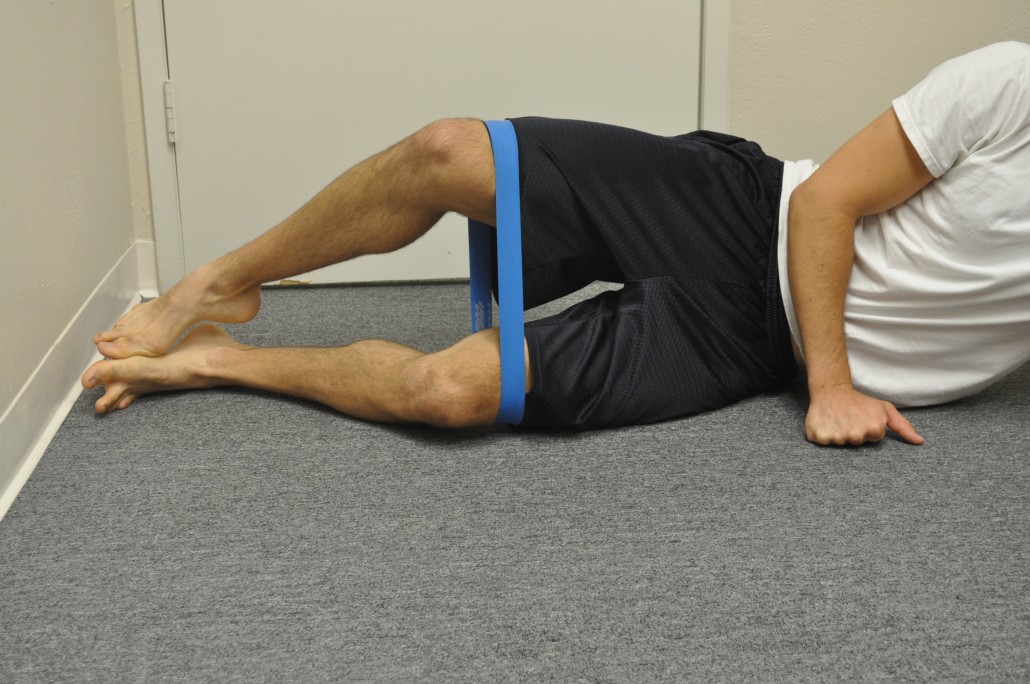
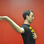
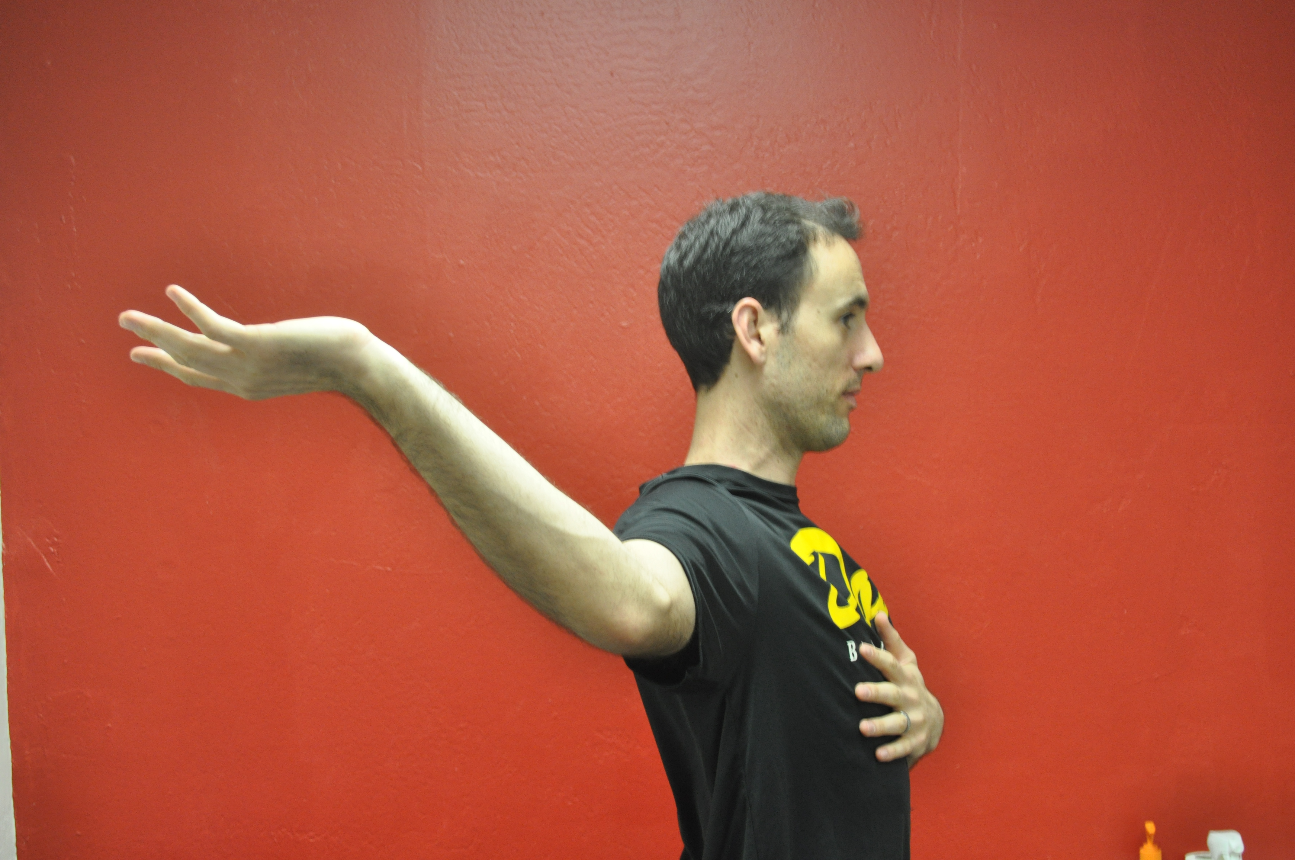
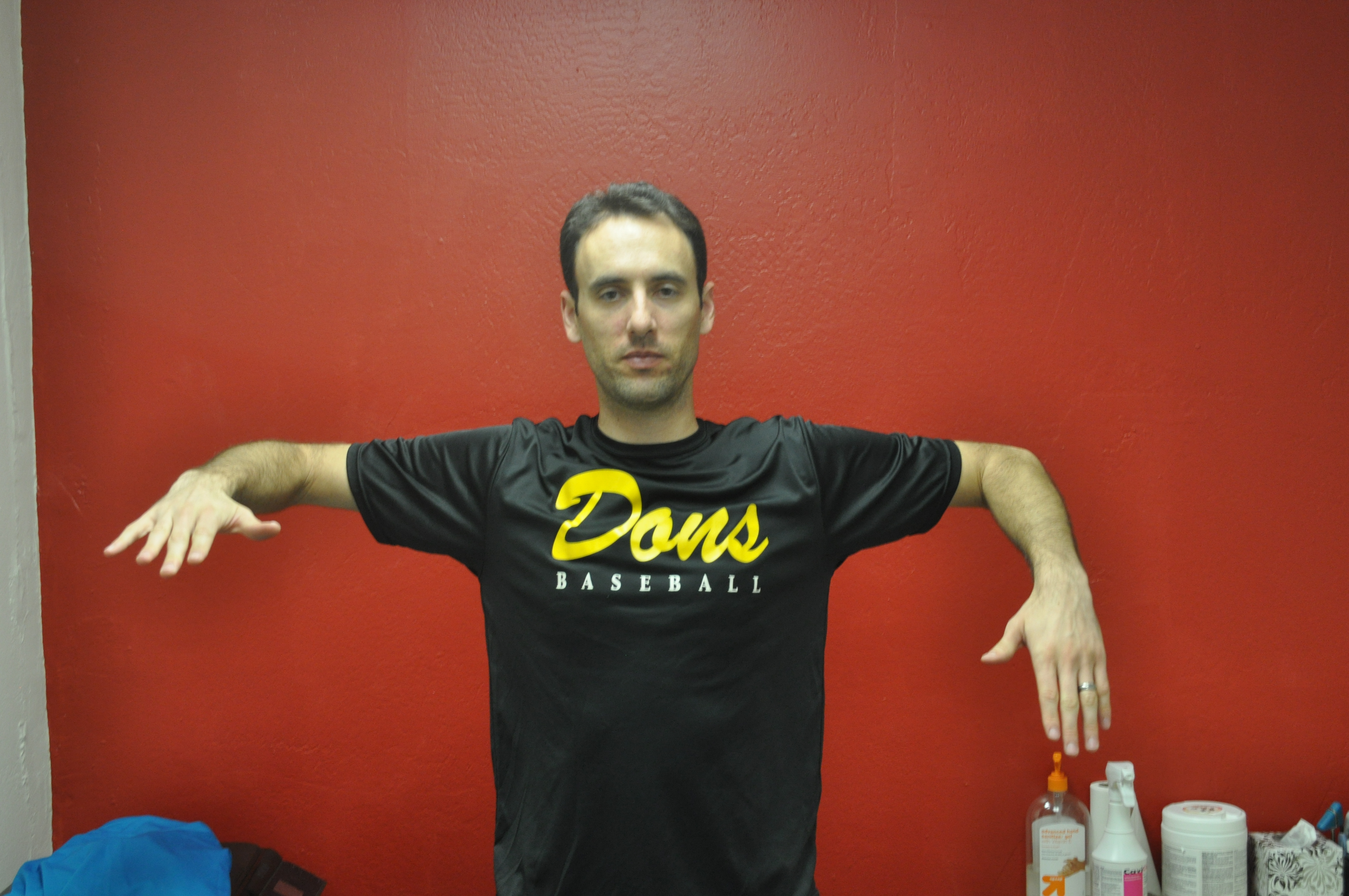
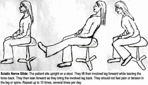
Raise knee as high as possible without losing positioning.

For runners, once pain has been minimal for 2 weeks and strengthening exercises are performed, a running routine can resume.3,4 Running should take place every other day on a flat surface with easy sprints and no hills.3,4 Gradual increases in frequency and duration of running should occur over a period for 3 to 4 weeks.4

Other Treatment Options

Other treatment options for ITBS include NSAIDs for pain and swelling.In chronic cases that have not responded to conservative care, corticosteroid injections may be warranted.10 In rare cases surgery may be performed to release the tension of the IT band over the lateral knee.11

Conclusion

If you are experiencing knee pain, it is important to get evaluated by a sports chiropractor to determine the exact cause. ITBS is a very common condition affecting approximately 8.4% of injured runners.9There are a number of conservative options for patients who are affected with this condition, particularly Active Release Technique (ART®) to help relieve pain associated with ITBS. ART®, chiropractic adjustments, stretching of the TFL/IT band, followed by gluteus medius strengthening and a gradual return to activity will result in most patients recovering from ITBS in 6 weeks.4


References

Meyers, T. W. (2009) Anatomy Trains: Myofascial Meridians for Manual and Movement Therapists. New York, NY: Elsevier.

Fredericson, M., and Weir, A. (2006). Practical management of iliotibial band friction syndrome in runners. Clinical Journal of Sports Medicine, 16 (3), 261-268.

Ellis, R., Hing, W., and Reid, D. (2007). Iliotibial band friction syndrome- a systematic review.  Manual Therapy, 12, 200-208.

Fredericson, M., Wolf, C. (2005). Iliotibial band syndrome in runners innovations in treatment. Sports Medicine, 35 (5), 451-459.

Fairclough, J. et al. (2006). The functional anatomy of the iliotibial band during flexion and extension of the knee: implications for understanding iliotibial band syndrome. Jounral of Anatomy, 208, 309-316.

Miller, R. H., Lowry, J. L., Meardon, S. A., and Gillette, J. C. (2006). Lower extremity mechanics of iliotibial band syndrome during an exhaustive run. Gait and Posture, 26, 407-413.

Ferber, R., Noehren, B., Hamill, J., and Davis, I. (2010). Competitive female runners with a history of iliotibial band syndrome demonstrate atypical hip and knee kinematics. Journal of Orthopaedic & Sports Physical Therapy, 40 (2), 52-58.

Hanten, W. P., Olson, S. L., Butts, N. L., and Nowicki, A. L. (2000). Effectiveness of a home program of ischemic pressure followed by sustained stretch for treatment of myofascial trigger points. Physical Therapy, 80 (10), 997-1003.

Taunton, J. E., Ryan, M. B., Clement, D. B., McKenzie, D. C., Lloyd-Smith, D. R., and Zumbo, B. D. (2002). A retrospective case-control analysis of 2002 running injuries. British Journal of Sports Medicine, 36, 95-101.

Hong, J. H., and Kim J. S. (2013). Dignosis of iliotibial band friction syndrome and ultrasound guided steroid injection.The Korean Journal of Pain, 26 (4), 387-391.

Sangkaew, C. (2007). Surgical treatment of iliotibial band friction syndrome with the mesh technique. Archives of Orthopaesic and Trauma Surgery, 127, 303-306.

dr. rose

Preventing Injuries – Baseball Pitcher

img005Overhead throwing creates a variety of challenges for baseball players, especially pitchers. Due to the biomechanical consequences of repetitive throwing, pitchers are at increased risk for a variety of shoulder and elbow injuries such as shoulder impingement, labral tears, and ulnar collateral ligament tears. These injuries can often lead to long periods of recovery and, in some cases, surgery.

One of the biomechanical changes that pitchers undergo is a loss of internal (or forward rotation) rotation in their shoulder joint. With repetitive cocking of the shoulder backward, pitchers develop tightness in an area of the back of the shoulder joint called the posterior capsule. This tightness leads to a decrease in the internal rotation of the throwing shoulder as compared to the non-throwing shoulder as seen in the images below. This is called Glenohumeral Internal Rotation Deficit or GIRD. GIRD has been shown to be a risk factor for SLAP labral tears and UCL ligament tears. Both injuries typically require surgery and recovery time of 12-18 months.


One way to avoid these debilitating injuries is to prevent GIRD. The best way to prevent GIRD or reduce GIRD, if it is already present, is through a simple stretch called the sleeper stretch. The sleeper stretch is performed by lying on your throwing side with your arm at 90 degrees from the trunk. From this position, the arm is rotated forward into internal rotation and held for 30 seconds (see images below). This stretch should be built into a pitcher’s daily routine. Over time, consistent stretching can lead to reversal of GIRD and thus a decrease in a significant risk factor for serious pitching injuries.


breast cancer survivor

Medieval Tools Increase Quality of Life of Breast Cancer Survivors

The Graston Technique for Breast Cancer Survivors

The Graston Technique is a form of deep tissue massage that is becoming increasingly popular. In the past, this method has proved to be effective for athletes as they overcome sport injuries, being utilized in the NBA and NFL. Scar tissue develops around an athlete’s injury causing chronic pain and a lack of mobility. Through the use of medieval looking stainless steel tools, the professionals are able to first locate the scarring as the equipment vibrates over hardened tissue. Then, they are able to manually break up the deep tissue, allowing the blood to flow freely. This removes the damaged tissue enabling the new tissue to grow.

More recently, the Graston Technique has proved to be successful in helping not only athletes but breast cancer patients as well. Whether the breast cancer survivor had a mastectomy or their lymph’s removed, this method is able to decrease pain and increase the individual’s range of motion and good posture. Chiropractors target the area of the surgery and are able to decrease the scarring through putting pressure on the deep, damaged tissues repetitively.

The method is painful, dissuading the faint of heart as it can lead to bruising. But, it’s long term effects are worth the temporary discomfort as it enables the athlete and the breast cancer survivor to have a better quality of life. If looking into it, the Graston Technique is not fully covered by insurance but the process is frequently a normal component of physical therapy.

whiplash san diego

Chiropractic and Whiplash – San Diego

Whiplash is the medical name for an injury to the neck that is created from a sudden jolt from front to back that creates a whip style movement. Whiplash is more often sustained in motor vehicle accidents; however, it can occur from a fall or sports/work related injury and so forth. The Whiplash injuries are normally due to the result of a sprain-strain to the neck, where the ligaments which provide support, protection and also limit the movement of the vertebrae are damaged. The most commonly injured joints are the facet joints which are located in the back portion of the spine. However, these are usually not the only injuries.

With more severe whiplash injuries tendons and muscles are strained and stretched, vertebral discs can be bulge or herniate, and the nerves may also suffer stretching and become irritated and inflamed. The most common symptoms that are felt with whiplash are stiffness and pain through the neck, generally found in the areas that have sustained damage. Most commonly pain will be in the front and back of the neck and turning the head will make the pain more severe. A headache is also a normal symptom of whiplash. Pain can also be found to extend through the upper part of the body.

In addition to the joint pain, some people experience dizziness, sickness, and even visual problems following a whiplash injury. These symptoms must not be ignored, and medical intervention should be sought if they do not resolve in a day or two. Whiplash symptoms are not always immediate and can take up to two days to appear.

Those suffering from whiplash need to stay active unless they have sustained an injury that requires immobilization. They may be worried but should move as much as possible. The doctor will more than likely prescribe some form of stretching exercises. These exercises are very important to aid recovery.

It is normal to use ice or heat to control the pain and reduce swelling after a whiplash injury. The injured party may also have electrical stimulation or ultrasound if necessary for short term relief. In the case of neck pain, spinal manipulation or spinal mobilization from a chiropractor can provide additional relief.

Active Release Technique

The Power of ART – Active Release Technique

Struggling with carpal tunnel can be a debilitating experience. The inflammation around tissues and nerves in the wrist can make it difficult to perform daily tasks. The classic symptoms of carpal tunnel include numbness or pain that occurs on the thumb-side of the hand, pain that radiates up to the shoulder, and the muscles in the thumb becoming severely distorted. However, there is no need to suffer with this condition when there is ART (Active Release Technique) to help you relieve the symptoms.

One can experience the relief of the pain and numbness without invasive surgery or traditional procedures of medicine. The continuance of these symptoms is the direct result of misdiagnosis and the misinterpretation of what carpal tunnel actually is. It’s more than just the entrapment of one single nerve in the thumb; rather, it’s a more common problem that takes place further up the arm, in the muscle called the Pronator Terres. Because of this, adding a brace to the wrist can actually make the problem worse.

ART, on the other hand, is designed to eliminate the problem from the get-go, allowing patients to make a much faster recovery than with conventional methods. Instead of focusing on just one area, ART aims to restore unimpeded range of motion and function to the soft tissues of the arm and wrist.

Professionals who are trained in the technique of ART can evaluate the texture and mobility of soft tissue, and, using hand pressure, removes or breaks up the fibrous adhesions that are present in the soft tissues. This can drastically improve the recovery from this debilitating condition, and abate the symptoms for much longer periods of time between each treatment.

There are typically three levels of ART that are performed by the practitioner himself while the fourth requires the patient to be involved with the active movement of the tissue while the practitioner applies the required tension to improve the results of the treatment even more. It has been scientifically proven that patients who are actively involved with the process of their treatment are more likely to make a better recovery than those who don’t.

Understand the true source of the pain you’re experiencing, and take steps to resolving your condition before it becomes too much to handle.

plantar fasciitis

Plantar Fasciitis: Resolving Foot Pain with Active Release Technique (ART) and Graston

Plantar Fasciitis is an extremely painful condition caused by inflammation and swelling of the plantar fascia, a thick band of tissue that begins at the back of your heel, runs across the underside of your foot. It often is caused by overuse, poor natural mechanics, excessive body weight, sports or improper footwear. Plantar fasciitis is one of the most common causes of foot pain. Some activities that can predispose a person are certain sports that involve running, ballet dancing, aerobics, improper stretching and even poor walking mechanics. Taut Achilles tendons, calf, hamstring, or weak foot muscles increase your risk factors. High arches or flat feet are natural biomechanics of your body that can contribute greatly to your chances of struggling with Plantar Fasciitis.

Symptoms

People with plantar fasciitis often complain of pain along the arch of the foot and at the heel. The pain can be excruciating when first applying pressure to the foot in the morning or after sitting or standing for long periods of time. The fascia supports the arch of the foot and becomes irritated and inflamed when pressure causes micro tearing of the tissues. The body’s response to this injury is to create scar tissue. The scar tissue is inelastic and can further perpetuate the problem by putting further mechanical strain on other areas of the foot creating a cycle of healing and injury. Plantar fasciitis can become a chronic injury, often termed plantar fasciosis at this point because there is minimal inflammation. Plantar fasciosis is often characterized by extensive scar tissue that restricts movement due to loss of blood flow to the area. It is very difficult to treat and can be nearly impossible to completely heal.

Home Care and Prevention

Check out our new blog post Top 7 Ways to Treat Plantar Fasciitis at Home. Every recommendation in that blog post are the current recommendations for patients to treat plantar fasciitis at home. Home care along with traditional treatment helps provide faster results!

Traditional Treatment

Common treatments used to alleviate pain and aid in the healing of plantar fasciitis are rest, ice, splints, orthotics, and non-steroidal anti-inflammatories.

Active Release Technique (ART) and Graston Technique

The primary cause of the pain in plantar fasciitis and plantar fasciosis is scar tissue adhesions, caused by repeated damage of the fascia. Traditional treatments alone or together may only minimally reduce the pain and do little to prevent recurrence.

ART and Graston Technique are relatively new approaches with very successful results. Being hands-on methods, the therapist can work directly with the soft tissues of the body. These treatments focus on locating and treating the scar tissues adhesions accumulated in the muscles and soft tissues.

The primary Goals of a practitioner using the ART and Graston techniques are to:

  1. Aid in the disruption/breakdown of the adhesions
  2. Increase normal tissue flexibility, movement and comfort
  3. Aim to restore full flexibility, balance and stability

Treatment of with ART and Graston techniques is applied to the plantar fascia, calf muscles, and other muscles of the foot. The treatment is complemented with specific foot strengthening and stability exercises. Most patients report a positive response within 2-4 treatments.

Sciatic Nerve Glide

Piriformis Syndrome Part III- Treatment

Piriformis Syndrome Treatment

The two main goals of treatment are 1) to relief irritation to the sciatic nerve and 2) address any functional problems that may be contributing.
If the sciatic nerve is inflamed the first step is to decrease aggravation from compression. Things to avoid are leg crossing, sitting on your wallet, and sitting on hard surfaces.  Things you should begin doing are applying ice to the piriformis, stretching of the piriformi, and taking oral anti-inflammatories (as directed).  Stretching every 2-3 hours (while awake) is a key to the success of non-operative treatment. Prolonged stretching of the piriformis muscle is accomplished in while lying on your back with the hip flexed and passively adducted/internally rotated (see picture).  Another exercise that can be helpful to decrease irritation between the piriformis and sciatic nerve is a nerve gliding exercise.  This exercise tensions the sciatic nerve and allows it to move relative to the piriformis. It is sometimes called nerve flossing to better describe how the nerve moves through other structures.  Two types of nerve gliding exercises are pictured below. (Pictures from http://www.dynamicchiropractic.com/mpacms/dc/article.php?id=53203)
 Sciatic Nerve GlideSciatic Nerve Tensioner
Manual therapy can be extremely helpful in releasing tension on the sciatic nerve and addressing functional deficits.  Soft tissue therapies, including myofascial release and Graston®, aimed at releasing tension and muscular adhesions within the piriformis can be beneficial.  Chiropractic spinal adjustments can help address functional contributions from the spine such as sacroiliac joint hypomobility and low back dysfunction.  Hyperpronation of the foot can be addressed through orthotics in the short term and foot strengthening in the long term.  Pronation is a normal foot motion however overpronation may lead to increased external rotation of the hip with walking putting tension on the piriformis and sciatic nerve.
Treatment may take several months of consistent work and it is important not to get discouraged. As long as there is no progression of neurological symptoms conservative treatment is the best option. Your symptoms should be monitored by a healthcare practitioner to ensure there is no progression.
-Dr. Kevin Rose, DC
Q Angle

Piriformis Syndrome Part II- Evaluation

Clinical Diagnosis

Piriformis syndrome occurs most frequently between ages 40-60 and is more common in women than men.  Some reports suggest a 6:1 female-to-male ratio for piriformis syndrome; , possibly because of biomechanics associated with the wider quadriceps femoris muscle angle (ie, “Q angle”) in the pelvis of women. Reported incidence rates for piriformis syndrome among patients with low back pain vary widely, from 5% to 36%.

Q-Angle

The most common  symptom of patients with piriformis syndrome is increasing pain after sitting for longer than 15 to 20 minutes. Many patients complain of pain over the piriformis muscle (ie, in the buttocks), especially over the muscle’s attachments at the sacrum and medial greater trochanter. Symptoms, which may be of sudden or gradual onset, are usually associated with spasm of the piriformis muscle or compression of the sciatic nerve; these symptoms include radiating/shooting pain or tingling or numbness in the back of the thigh, leg, or foot.  These symptoms must be evaluated by a healthcare provider to differentiate the possible causes.  Patients may also complain of difficulty walking and of pain with internal rotation of the involved leg, such as occurs during cross-legged sitting or walking. X-rays or an MRI offer little help in directly diagnosing piriformis syndrome but may be used to rule out other causes of sciatica such as a herniated disc in the lumbar spine.

Functional Evaluation

There are many functional abnormalities that may have either caused or resulted from this condition.  Once the diagnosis has been made, these underlying, perpetuating biomechanical factors must be addressed.

Functional biomechanical deficits associated with piriformis syndrome may include the following:

  • Tight hip external rotators including pirifromis
  • Tight adductors (groin)
  • Hip abductor weakness
  • Lower lumbar spine dysfunction
  • Sacroiliac joint hypomobility
  • Hyperpronation of the foot and prolonged toe-off

Functional adaptations to these deficits include the following:

  • Ambulation with the thigh in external rotation
  • Functional limb length shortening
  • Shortened stride length

Next post will discuss treatment options for piriformis syndrome

Piriformis Syndrome

Piriformis Syndrome: Overview and Causes

Piriformis SyndromePiriformis syndrome is a unique cause if sciatic nerve irritation (neuritis) or sciatica. The condition, which can mimic lumbar disc herniation, usually is caused by irritation of the sciatic nerve due to spasm and/or contracture of the piriformis muscle. Piriformis syndrome is also referred to as “pseudosciatica”, “wallet sciatica”, and “hip socket neuropathy”.
It frequently goes unrecognized or is misdiagnosed in clinical settings. Piriformis syndrome can “masquerade” as other common somatic dysfunctions, such as intervertebral discitis, lumbar radiculopathy, primary sacral dysfunction, sacroiliitis, sciatica, and trochanteric bursitis.


(Image from http://www.concordortho.com/patient-education/topic-detail-popup.aspx?topicID=4214fc65d020761633286131e407d037)

Anatomical Considerations

The proper understanding of piriformis syndrome requires knowledge of the anatomy and anatomical variations in the relationships between the sciatic nerve and the piriformis muscle.

The piriformis muscle is flat, pyramid-shaped, and oblique. This muscle originates on the front of the sacrum and inserts at the greater trochanter of the femur. With the hip extended, the piriformis muscle is an external rotator of the hip; however, with the hip flexed, the muscle becomes a hip abductor.

In most of the population, the sciatic nerve exits the pelvis deep along the lower surface of the piriformis muscle. However, many developmental variations of the relationship between the sciatic nerve in the pelvis and piriformis muscle have been observed. In as much as 22% of the population, the sciatic nerve pierces the piriformis muscle, splits the piriformis muscle, or both, predisposing these individuals to irritation of the sciatic nerve.

Sciatic N and Piriformis Orientation


From http://www.anatomyatlases.org/AnatomicVariants/NervousSystem/Images/70.shtml

Causes of Piriformis Syndrome

Piriformis syndrome can be caused by a variety of issues. The underlying mechanism is from irritation to the sciatic nerve. Below are some causes of irritation to the sciatic nerve as it passes the piriformis muscle:

1. Muscular problems

  • Spasms and adhesions in the piriformis muscle cause compression and irritation of the sciatic nerve. Muscular damage or tightness can develop from a single injury or repetitive use injury. Vigorous physical activity can lead to such an injury- (commonly seen in athletes such as runners, cyclists, and dancers).

2. Postural

  • Hyperlordosis (increased curvature of the low back) and increased foot pronation are both risk factors for piriformis syndrome

3. Traumatic

  • Direct compression of the piriformis and/or sciatic nerve from an external soure such as a wallet.

4. Partial or total nerve anatomical abnormalities

  • An anomaly in the nerve itself as it passes through the piriformis muscle can lead to dysfunction

5. Other causes can include the following:

  • Pseudoaneurysms of the inferior gluteal artery adjacent to the piriformis syndrome
  • Bilateral piriformis syndrome due to prolonged sitting during an extended neurosurgical procedure
  • Cerebral Palsy
  • Total hip arthroplasty
  • Myositis ossificans

Stay tuned…… the next post will talk about how this problem is diagnosed and treated.