Sports injuries are injuries that occur in athletic activities. They can result from acute trauma, or from overuse of a particular body part

Preventing Soccer Injuries

Preventing Soccer Injuries

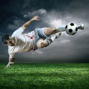
Soccer is an excellent way to build endurance, improve speed and stay in shape, all while enjoying being a part of a team. Nevertheless, soccer does involve quick start-and-stop motions and physical contact, which can lead to injury. The risk of injury is no reason not to play soccer however. Players simply need to be aware of the risks and know what steps they can take to play as safely as possible.

Sprains and strains are very common injuries in soccer, with varying forms of severity. Other injuries could include stress fractures and muscle strains from repeated overuse and direct blows to the body. Soccer players are also prone to shin splints, patellar tendinitis, and Achilles tendinitis. These injuries occur in the lower extremities, whereas neck sprains and concussions are common in the upper extremities. Injuries to the head, neck face are sometimes unavoidable. Just like wrist sprains, fractures and shoulder dislocations are unfortunately common due to falls and player-to-player contact.

The best place to start in prevention of these injuries is to have a pre-season physical examination and follow your doctor’s recommendations during the season. Using well-fitting cleats and shin guards is important in prevention. Molded and multi-studded cleats may be safer than screw-in cleats. Stay hydrated when out on the field and maintain proper fitness throughout the sport’s season. Injury rates are known to be higher in athletes who have not adequately prepared physically and remember to stretch adequately before and after your time on the field. After being inactive in the sport, progress gradually back to full-contact soccer through activities such as aerobic conditioning, agility training, and strength training.

Some of the greatest advice in preventing soccer injuries is to avoid overuse injuries. When it comes to the health of your body, more is not always better. Many sports medicine experts believe that it is helpful to take at least one season off each year. Although it’s hard, try to avoid the pressure that is now forced on many young athletes to over-train. Always listen to your body and decrease training time and intensity if pain or discomfort continues to increase. It will reduce the risk of injury and help avoid “burn-out.” Most injuries can be prevented to an extent. The last thing anyone wants to do is put an early end to his/her soccer career because they didn’t listen to their body and take care of themselves. As always, speak with a sports medicine professional if you have any concerns about injuries or soccer injury prevention strategies.

In order to keep kids and adults out on the field long-term and enjoy the sport of soccer, injury prevention, early detection, and treatment are significantly important.

golf injury san diego

Golf is a Game of Recovery

Golf is a sport that takes patience, diligence, and precision. Priority is not placed on the max a golfer can bench (their strength) or the pace at which they can run (their speed and endurance) like a majority of other sports. It is in the ability of the golfer to allow the club to do the work, to relax in their stance, and keep their eyes on the ball. Golf takes practice. The best golfers start at a young age and practice, practice, and practice. This is the reason that one of the greatest causes of injuries in golfers is overuse.

Overuse injuries occur over time. They are caused by repetitive motion. The most common overuse injuries for golfers are injuries in the joints, muscles, and tendons of a golfer’s back, shoulders, and elbows. Studies show that 44 percent of injuries in youth golfers are due to overuse. Children and adults are more prone to injury if they are not flexible, in bad condition, practice and play an extreme amount, do not use the correct form, or hit the ground often. Studies have indicated that the number of times a golfer hits a ball is directly correlated to the number of injuries that golfer has.

Acute injuries generally occur when a golfer does not use the correct technique. It is an injury that happens at one moment, but takes days, weeks, or even months to recover from. A golfer using the wrong form, hitting the ground too hard, or carrying their bag incorrectly can cause this type of injury.

The greatest factor causing golfers to face overuse or acute injuries is a lack of flexibility. Studies show that the majority of golfers (80%) take less than 10 minutes to warm up and stretch before a round of golf. Those who did not warm up had double the amount of injuries than those who did. This is also the easiest, most practical way for a golfer to prevent injury. Increase flexibility and decrease the amount of injuries. A golfer should also ensure that their techniques are correct. If their form is wrong, they greatly increase their risk of injury.

dr. rose

Preventing Injuries – Baseball Pitcher

img005Overhead throwing creates a variety of challenges for baseball players, especially pitchers. Due to the biomechanical consequences of repetitive throwing, pitchers are at increased risk for a variety of shoulder and elbow injuries such as shoulder impingement, labral tears, and ulnar collateral ligament tears. These injuries can often lead to long periods of recovery and, in some cases, surgery.

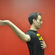
One of the biomechanical changes that pitchers undergo is a loss of internal (or forward rotation) rotation in their shoulder joint. With repetitive cocking of the shoulder backward, pitchers develop tightness in an area of the back of the shoulder joint called the posterior capsule. This tightness leads to a decrease in the internal rotation of the throwing shoulder as compared to the non-throwing shoulder as seen in the images below. This is called Glenohumeral Internal Rotation Deficit or GIRD. GIRD has been shown to be a risk factor for SLAP labral tears and UCL ligament tears. Both injuries typically require surgery and recovery time of 12-18 months.


One way to avoid these debilitating injuries is to prevent GIRD. The best way to prevent GIRD or reduce GIRD, if it is already present, is through a simple stretch called the sleeper stretch. The sleeper stretch is performed by lying on your throwing side with your arm at 90 degrees from the trunk. From this position, the arm is rotated forward into internal rotation and held for 30 seconds (see images below). This stretch should be built into a pitcher’s daily routine. Over time, consistent stretching can lead to reversal of GIRD and thus a decrease in a significant risk factor for serious pitching injuries.


breast cancer survivor

Medieval Tools Increase Quality of Life of Breast Cancer Survivors

The Graston Technique for Breast Cancer Survivors

The Graston Technique is a form of deep tissue massage that is becoming increasingly popular. In the past, this method has proved to be effective for athletes as they overcome sport injuries, being utilized in the NBA and NFL. Scar tissue develops around an athlete’s injury causing chronic pain and a lack of mobility. Through the use of medieval looking stainless steel tools, the professionals are able to first locate the scarring as the equipment vibrates over hardened tissue. Then, they are able to manually break up the deep tissue, allowing the blood to flow freely. This removes the damaged tissue enabling the new tissue to grow.

More recently, the Graston Technique has proved to be successful in helping not only athletes but breast cancer patients as well. Whether the breast cancer survivor had a mastectomy or their lymph’s removed, this method is able to decrease pain and increase the individual’s range of motion and good posture. Chiropractors target the area of the surgery and are able to decrease the scarring through putting pressure on the deep, damaged tissues repetitively.

The method is painful, dissuading the faint of heart as it can lead to bruising. But, it’s long term effects are worth the temporary discomfort as it enables the athlete and the breast cancer survivor to have a better quality of life. If looking into it, the Graston Technique is not fully covered by insurance but the process is frequently a normal component of physical therapy.

whiplash san diego

Chiropractic and Whiplash – San Diego

Whiplash is the medical name for an injury to the neck that is created from a sudden jolt from front to back that creates a whip style movement. Whiplash is more often sustained in motor vehicle accidents; however, it can occur from a fall or sports/work related injury and so forth. The Whiplash injuries are normally due to the result of a sprain-strain to the neck, where the ligaments which provide support, protection and also limit the movement of the vertebrae are damaged. The most commonly injured joints are the facet joints which are located in the back portion of the spine. However, these are usually not the only injuries.

With more severe whiplash injuries tendons and muscles are strained and stretched, vertebral discs can be bulge or herniate, and the nerves may also suffer stretching and become irritated and inflamed. The most common symptoms that are felt with whiplash are stiffness and pain through the neck, generally found in the areas that have sustained damage. Most commonly pain will be in the front and back of the neck and turning the head will make the pain more severe. A headache is also a normal symptom of whiplash. Pain can also be found to extend through the upper part of the body.

In addition to the joint pain, some people experience dizziness, sickness, and even visual problems following a whiplash injury. These symptoms must not be ignored, and medical intervention should be sought if they do not resolve in a day or two. Whiplash symptoms are not always immediate and can take up to two days to appear.

Those suffering from whiplash need to stay active unless they have sustained an injury that requires immobilization. They may be worried but should move as much as possible. The doctor will more than likely prescribe some form of stretching exercises. These exercises are very important to aid recovery.

It is normal to use ice or heat to control the pain and reduce swelling after a whiplash injury. The injured party may also have electrical stimulation or ultrasound if necessary for short term relief. In the case of neck pain, spinal manipulation or spinal mobilization from a chiropractor can provide additional relief.

Active Release Technique

The Power of ART – Active Release Technique

Struggling with carpal tunnel can be a debilitating experience. The inflammation around tissues and nerves in the wrist can make it difficult to perform daily tasks. The classic symptoms of carpal tunnel include numbness or pain that occurs on the thumb-side of the hand, pain that radiates up to the shoulder, and the muscles in the thumb becoming severely distorted. However, there is no need to suffer with this condition when there is ART (Active Release Technique) to help you relieve the symptoms.

One can experience the relief of the pain and numbness without invasive surgery or traditional procedures of medicine. The continuance of these symptoms is the direct result of misdiagnosis and the misinterpretation of what carpal tunnel actually is. It’s more than just the entrapment of one single nerve in the thumb; rather, it’s a more common problem that takes place further up the arm, in the muscle called the Pronator Terres. Because of this, adding a brace to the wrist can actually make the problem worse.

ART, on the other hand, is designed to eliminate the problem from the get-go, allowing patients to make a much faster recovery than with conventional methods. Instead of focusing on just one area, ART aims to restore unimpeded range of motion and function to the soft tissues of the arm and wrist.

Professionals who are trained in the technique of ART can evaluate the texture and mobility of soft tissue, and, using hand pressure, removes or breaks up the fibrous adhesions that are present in the soft tissues. This can drastically improve the recovery from this debilitating condition, and abate the symptoms for much longer periods of time between each treatment.

There are typically three levels of ART that are performed by the practitioner himself while the fourth requires the patient to be involved with the active movement of the tissue while the practitioner applies the required tension to improve the results of the treatment even more. It has been scientifically proven that patients who are actively involved with the process of their treatment are more likely to make a better recovery than those who don’t.

Understand the true source of the pain you’re experiencing, and take steps to resolving your condition before it becomes too much to handle.

plantar fasciitis

Plantar Fasciitis: Resolving Foot Pain with Active Release Technique (ART) and Graston

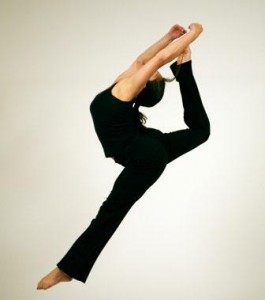
Plantar Fasciitis is an extremely painful condition caused by inflammation and swelling of the plantar fascia, a thick band of tissue that begins at the back of your heel, runs across the underside of your foot. It often is caused by overuse, poor natural mechanics, excessive body weight, sports or improper footwear. Plantar fasciitis is one of the most common causes of foot pain. Some activities that can predispose a person are certain sports that involve running, ballet dancing, aerobics, improper stretching and even poor walking mechanics. Taut Achilles tendons, calf, hamstring, or weak foot muscles increase your risk factors. High arches or flat feet are natural biomechanics of your body that can contribute greatly to your chances of struggling with Plantar Fasciitis.

Symptoms

People with plantar fasciitis often complain of pain along the arch of the foot and at the heel. The pain can be excruciating when first applying pressure to the foot in the morning or after sitting or standing for long periods of time. The fascia supports the arch of the foot and becomes irritated and inflamed when pressure causes micro tearing of the tissues. The body’s response to this injury is to create scar tissue. The scar tissue is inelastic and can further perpetuate the problem by putting further mechanical strain on other areas of the foot creating a cycle of healing and injury. Plantar fasciitis can become a chronic injury, often termed plantar fasciosis at this point because there is minimal inflammation. Plantar fasciosis is often characterized by extensive scar tissue that restricts movement due to loss of blood flow to the area. It is very difficult to treat and can be nearly impossible to completely heal.

Home Care and Prevention

Check out our new blog post Top 7 Ways to Treat Plantar Fasciitis at Home. Every recommendation in that blog post are the current recommendations for patients to treat plantar fasciitis at home. Home care along with traditional treatment helps provide faster results!

Traditional Treatment

Common treatments used to alleviate pain and aid in the healing of plantar fasciitis are rest, ice, splints, orthotics, and non-steroidal anti-inflammatories.

Active Release Technique (ART) and Graston Technique

The primary cause of the pain in plantar fasciitis and plantar fasciosis is scar tissue adhesions, caused by repeated damage of the fascia. Traditional treatments alone or together may only minimally reduce the pain and do little to prevent recurrence.

ART and Graston Technique are relatively new approaches with very successful results. Being hands-on methods, the therapist can work directly with the soft tissues of the body. These treatments focus on locating and treating the scar tissues adhesions accumulated in the muscles and soft tissues.

The primary Goals of a practitioner using the ART and Graston techniques are to:

  1. Aid in the disruption/breakdown of the adhesions
  2. Increase normal tissue flexibility, movement and comfort
  3. Aim to restore full flexibility, balance and stability

Treatment of with ART and Graston techniques is applied to the plantar fascia, calf muscles, and other muscles of the foot. The treatment is complemented with specific foot strengthening and stability exercises. Most patients report a positive response within 2-4 treatments.

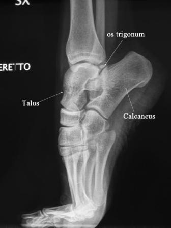
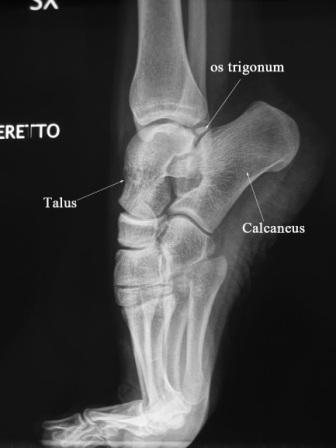
Lateral Demipointe Compressed - Xray of Ankle

POSTERIOR IMPINGEMENT IN DANCERS

Posterior ankle impingement is a common cause of pain in ballet dancers.   Other names for this condition are “os trigonum syndrome” and “nutcracker syndrome”.  It is called “nutcracker syndrome” not because of its common occurrence in ballet at Christmas-time but because of the way the tissue of the ankle is squeezed at the ankle.  The following is a brief overview of this condition and how it relates to dancers.

Posterior impingement of the ankle is often attributable to the presence of an accessory bone growth called an os trigonum or a Stieda process that is located just behind the talus (see x-ray for anatomy).   The stress from repetitive plantarflexion by dancers, especially at a young age, is the suspected cause of the development of the os trigonum.  Pain in posterior impingement occurs when the soft tissue of the ankle is pinched between the posterior lip of the tibia and the calcaneus.  This occurs when the foot is in extreme plantarflexion such as during releve in the demipointe or en pointe positions.

Diagnosis
Ankle pain ballet San Diego

Proper diagnosis of posterior impingement is imperative for recovery from this injury.  Posterior impingement attributable to an os trigonum is usually misdiagnosed as Achilles’ tendonitis/tendonopathy, peroneal tendonitis or flexor hallucis longus strain.

The two main symptoms of posterior impingement are a decrease in plantarflexion compared with the unaffected ankle and pain in the posterior region of the ankle.  Often dancers are aware of a lack of ability to fully pointe in one foot compared to the other, this may be an early sign of impingement.   Another common description dancer’s use is it feels like pinching in the heel during releve.  Diagnosis is often aided by x-Rays of the ankle.  It is best to request an x-ray to be taken during releve to evaluate the biomechanics of the injury.  If there has been persistent pain for a period of 1– 4 months, local swelling, and radiographic assessment indicating a posterior ankle impingement, then an MRI should be performed.

Treatment for Posterior Impingement of the Ankle

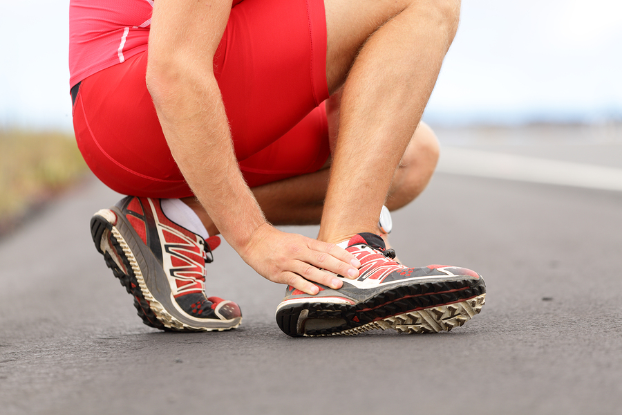
Once posterior impingement is diagnosed focused treatment should begin.  Non-surgical care is usually successful and should be the first line treatment.  Treatment should be focused on taking pressure off of the tissue being pinched.  Exercises should focus on engaging the deep muscles of the leg especially the deep flexors.  One exercise that is helpful and can be done at home is a self traction maneuver with plantarflexion (see picture).  The patient holds his or her ankle, as shown, with downward pressure and performs the motion with a bent knee.  Bending the knee helps disengage the gastrocnemius muscle and soleus forcing the deep flexors to engage. Post Impingement Exercise Compressed Another great exercise is ankle range of motion with traction applied by a therapist using very strong elastic bands.  Dancers may experience relief with traction and feel they are able to fully plantarflex; this can also be a good way to support the diagnosis as Achilles’ tendonitis is often unchanged with traction.  Manipulation of the ankle especially the talus can provide relief as well. Conservative therapy is successful in the majority of cases. Recovery may take several months.

A surgical approach should only be adopted in the following cases:

  • recurrent or unremitting symptoms in professional ballet dancers;
  • persistent decreased plantarflexion compared with the unaffected ankle;
  • failure of physical and medical therapies after 1– 4 months (depending on the level of the athlete/dancer);
  • posterior impingement clinically suspected and indicated by both x-ray and MRI.

Ankle pain and heel pain is a common symptom in dancers and posterior impingement is only one of the causes. If you think you may be suffering from posterior impingement seek advice from a qualified healthcare professional with expertise in dance injury.

-Dr. Rose, DC, CCSP®

Dr. Rose is a Certified Chiropractic Sports Practitioner® with experience in dance medicine.  He is currently Director of Physical Rehabilitation at Ballet San Jose and a member of the International Association of Dance Medicine and Science.

REFERENCES

Albisetti W, Ometti M, Pascale V, De Bartolomeo O: Clinical evaluation and treatment of posterior impingement in dancers. Am J Phys Med Rehabil 2009;88:349–354.

Niek van Dijk C: Anterior and posterior ankle impingement. Foot Ankle Clin 2006;11:663– 83

F Cilli, M Akcaoglu: The incidence of accessory bones of the foot and their clinical significance. Acta Orthop Traumatol Turc 2005;39:243– 6

Stretching Aerobics

POSTURAL STABILITY IN DANCERS AFTER INJURY

Dancer in ActionBallet dancers are widely known for their superior body control in various challenging body positions.  In fact it has been found in recent studies that ballet dancers have better postural control when compared to other elite athletes. However, what has not been examined is the effect of injury on postural balance in dancers.  A recent study in The American Journal of Sports Medicine by Lin, et al, addresses this vary issue.

Postural balance requires a combination of several different sensory inputs: visual, vestibular (inner ear), and somatosensory (from the skin, joints, muscles, etc). Somatosensory input gives the brain information about where the body is in space; this is called proprioception.  These sensors can be damaged with ligamentous and muscular injuries such as ankle sprains and in turn lead to a deficit in proprioception.  Proprioceptive deficits can put a dancer at more risk for re-injury or new injury.

The recent study by Lin was the first study that evaluated stability in dancers in ballet specific postures following an injury. Three different groups were studied: healthy dancers, injured dancers (with sprained ankles), and health non-dancers.  Postural stability was evaluated on a balance sensor that monitors subtle deviations in stability.  This was done with the eyes open and then again with the eyes closed to further stress the proprioceptive component of balance.  As expected the injured dancers had decreased control not only in ballet specific positions but also in a simple single leg standing position when compared to healthy dancers. Surprisingly the injured dancers had worse stability control than the non-dancers.  Many dancers, and other athletes, that I have treated often assume that a skill such as balancing will come back without training, however this is not the case, and as the study indicated it regresses to the point that is worse than untrained people.Ankle

The postural deficits were most notable in side to side control with single leg standing and front to back control with first and fifth position.  Compensation was noted at the knee and hip joints in an attempt to make up for the injured ankle.  The en pointe position crated the most stability issues; injured dancers were inferior in all directions of stability.  With lateral ankle sprains the most commonly damaged ligament is the anterior talofibular ligament. This ligament is stressed maximally when the ankle is plantar flexed, such as during en pointe positions.  It is not surprising that when this ligament is damaged the proprioception during pointe is significantly disturbed.

This study brings to light the importance of a thorough rehabilitation program that includes proprioceptive training in ballet specific positions.  Typically a sprained ankle takes 6-8 weeks to recover.  During this time it is critical to begin proprioceptive training to ensure that the balance deficits do not lead to further injury.  Additionally if a dancer has a past injury of ankle sprain without proper rehabilitation it is likely that postural stability still remains and there is an increased risk to further injury.  If this is the case, dancers should begin a 3-4 week proprioceptive training plan that includes ballet specific posture.

Dance injuries and rehabilitation are unique and should be managed by a healthcare practitioner that is experienced with the subtleties of dance medicine.  Proprioception training for ballet typically includes balance exercises on an unstable surface such as a Bosu ball or stability disc.  These exercises should be done in normal stance position as well as ballet specific positions.  Whole body vibration may additionally add value as a proprioceptive trainer especially early during rehab when range of motion is limited.  KinesioTape or RockTape are also good taping methods that help protect the joint and provide increased proprioception when applied properly. Keep in mind that a training plan should be designed specifically for each injury and you should not attempt to manage the injury without the supervision of an experienced healthcare practitioner.

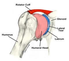
Glabrum - Picture of Shoulder

Labral tears of the shoulder: surgery or not?


At the recent American Orthopaedic Society for Sports Medicine, a comparison of non-surgical treatment and surgical treatment for SLAP labral tears of the shoulder in baseball players was presented. The labrum is a cartilage structure that makes the Glabrum - Labral Shoulder Tearsocket of the shoulder deeper and helps hold the upper arm. SLAP tears are a specific type of labral injury that is mainly caused by repetitive throwing.   Although surgical repair of SLAP tears is common, little is known about the effectiveness of nonsurgical pain relief and the effects of surgery on performance.

Return to play

Return to same level of play

Surgery

48%

7%

Non-Surgery

40%

22%


The return to play rate in SLAP injuries is low, typically reported at less than 50%. In this study the return to play rate for those that underwent surgery was 48% while those that underwent rehabilitation was only slightly lower at 40%. The most interesting finding in this study however was that the group that had surgery rarely, only 7% of the time, returned to the level of play that they had previously attained. The non-surgery group in comparison returned to the same level of play 22% of the time.

John E. Kuhn, MD, associate professor of orthopedic surgery and rehabilitation and chief of shoulder surgery at Vanderbilt University Medical Center in Nashville, Tennessee described the findings as “very significant” and summed up the findings:

“A lot of these throwing athletes can be treated non-operatively,” Dr. Kuhn said. “They had very good success with rehabilitation.  That suggests that patients can throw with SLAP tears, and that not every SLAP tear needs to be repaired,” he said. “Many can be rehabilitated; the sources of pain or disability may not even be the SLAP lesion itself,” he added. The research suggests that surgery in this patient population really should be a career-salvaging option, he explained. “It really shouldn’t be something you throw at somebody quickly or early. Rehab them, do everything you can to prevent surgery,” he concluded.

Another take home message from these findings that should be discussed further is the importance of avoiding shoulder injuries from throwing.  As stated the percentage of those injured with SLAP tears that return to play is less than 50%.  The best advice then logically is to avoid getting a SLAP tear altogether.  Unfortunately, there is no sure fire way to avoid getting a tear if you are a pitcher. However what can be done is limiting the risk factors for injury.  Risk factors for throwing injuries include a decrease in shoulder internal rotation, poor scapular control, and high pitching load.  These risk factors should be screened for and proper preventative exercises should be built into any strength and conditioning program for baseball players, especially pitchers. Non surgical treatment in these cases included rehabilitation that focused on stretching the posterior capsule of the shoulder to increase internal rotation and training the scapular muscles to hold the scapula in a stable position during the wind-up and cocking phase of the throwing motion. These exercises should also be at the core of any preventative arm care program for baseball players. Manual therapy, including myofascial release and Graston treatment can also be beneficial to aid in increasing range of motion.  These preventative strategies should be overseen by a healthcare provider knowledgeable in baseball injuries.

If you would like more information about how to recover from or prevent shoulder injuries contact our office today.

Another article outlining preventative exercises is coming soon…

-Dr. Kevin Rose, DC


 References

Yin, S.”SLAP Tears Often Treated Successfully without Surgery”. Medscape Medical News, July 7 2012.

Wilk et al. Passive range of motion characteristics in the overhead baseball pitcher and their implications for rehabilitation.  Clin Orthop Relat Res. 2012 Jun;470(6):1586-94.

Wilk et al. Shoulder injuries in te overhead athlete.  J Orthop Sports Phys Ther. 2009 Feb;39(2):38-54.